
The contemporary role of coronary physiology methods and intravascular imaging in percutaneous treatment of coronary artery disease
Article Sidebar

This work is licensed under a Creative Commons Attribution-ShareAlike 4.0 International License.
Main Article Content
 https://orcid.org/0000-0002-8133-9318
Abstract
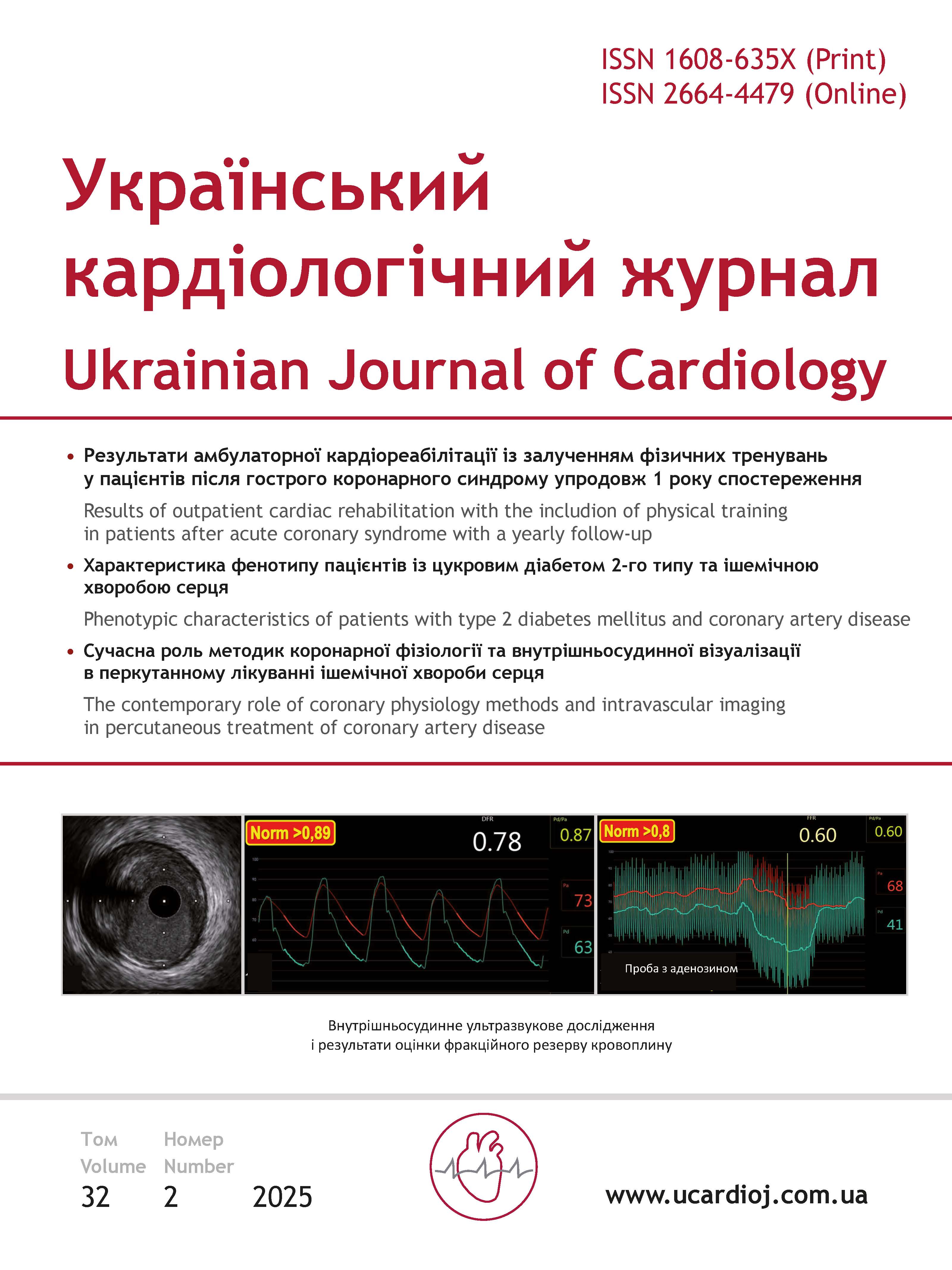
Ischemic heart disease (IHD) remains the leading pathology among cardiovascular diseases, showing a trend of increasing morbidity and a younger age of patients. The incidence of IHD grows every year, with patients becoming younger and mortality rates rising. Without timely diagnosis and effective treatment, this can lead to catastrophic consequences. IHD occurs due to an imbalance between the heart muscle’s oxygen demand and its delivery through the coronary blood flow. Timely diagnosis and effective treatment of IHD are critically important to prevent catastrophic events such as myocardial infarction or sudden cardiac death. The main task is to detect atherosclerotic lesions and determine their severity. Modern visualization methods, such as intravascular ultrasound (IVUS) and fractional flow reserve (FFR), are highly reliable invasive methods for assessing the potential for ischemic damage, with an accuracy of over 90 %. These methods are especially useful for evaluating intermediate coronary lesions in stable ischemic heart disease. Therefore, timely diagnosis and the use of modern methods for assessing atherosclerotic lesions are key to effective treatment of IHD and preventing serious complications. This article discusses the significance of visualization methods, supported by international multicenter studies such as DEFER, FAME-1, FAME-2, and FAME-3. In the presented clinical case of a patient with intermediate coronary artery stenosis, an evaluation was performed using fractional flow reserve (FFR) and intravascular ultrasound (IVUS). FFR helps determine the functional significance of a stenosis by measuring pressure before and after the narrowing, which assists in deciding whether revascularization is necessary. IVUS provides detailed visualization of the vessel’s internal structure, allowing the assessment of plaque morphology and the degree of stenosis.
Article Details
Keywords:
References
Vaduganathan M, Mensah George A, Turco Justine V, Fuster V, Roth Gregory A. The Global Burden of Cardiovascular Diseases and Risk. J Amer Coll Cardiol. 2022;80(25):2361-71. https://doi.org/10.1016/j.jacc.2022.11.005
Mensah GA, Fuster V, Murray CJL, Roth GA, et al. Global Burden of Cardiovascular Diseases and Risks, 1990-2022. J Amer Coll Cardiol. 2023;82(25):2350-473. https://doi.org/10.1016/j.jacc.2023.11.007
Shah SN. Coronary Artery Atherosclerosis: Emedicine; 2023 [updated Jul 20, 2023] https://doi.org/10.1161/CIR.0000000000001168
Libby P, Theroux P. Pathophysiology of Coronary Artery Disease. Circulation. 2005;111(25):3481-8. https://doi.org/10.1161/circulationaha.105.537878
Samady H, Eshtehardi P, McDaniel MC, Suo J, Dhawan SS, Maynard C, et al. Coronary Artery Wall Shear Stress Is Associated With Progression and Transformation of Atherosclerotic Plaque and Arterial Remodeling in Patients With Coronary Artery Disease. Circulation. 2011;124(7):779-88. https://doi.org/10.1161/circulationaha.111.021824
Kolodgie FD, Gold HK, Burke AP, Fowler DR, Kruth HS, Weber DK, et al. Intraplaque Hemorrhage and Progression of Coronary Atheroma. New Engl J Med. 2003;349(24):2316-25. https://doi.org/10.1056/nejmoa035655
Garcìa-Garcìa HM, Gogas BD, Serruys PW, Bruining N. IVUS-based imaging modalities for tissue characterization: similarities and differences. The Intern J Cardiovasc Imaging. 2011;27(2):215-24. https://doi.org/10.1007/s10554-010-9789-7
Levine GN, Bates ER, Blankenship JC, Bailey SR, Bittl JA, Cercek B, et al. 2011 ACCF/AHA/SCAI guideline for percutaneous coronary intervention: a report of the American College of Cardiology Foundation/American Heart Association Task Force on Practice Guidelines and the Society for Cardiovascular Angiography and Interventions. Catheter Cardiovasc Interv. 2013;82(4):E266-355. https://doi.org/10.1161/cir.0b013e31823ba622
Fischer JJ, Samady H, McPherson JA, Sarembock IJ, Powers ER, Gimple LW, et al. Comparison between visual assessment and quantitative angiography versus fractional flow reserve for native coronary narrowings of moderate severity. Amer J Cardiol. 2002;90(3):210-5. https://doi.org/10.1016/s0002-9149(02)02456-6
McDaniel MC, Eshtehardi P, Sawaya FJ, Douglas JS, Samady H. Contemporary Clinical Applications of Coronary Intravascular Ultrasound. JACC: Cardiovascular Interventions. 2011;4(11):1155-67. https://doi.org/10.1016/j.jcin.2011.07.013
Secemsky EA, Parikh SA, Kohi M, Lichtenberg M, Meissner M, Varcoe R, et al. Intravascular ultrasound guidance for lower extremity arterial and venous interventions. EuroIntervention. 2022;18(7):598-608. https://doi.org/10.4244/eij-d-21-00898
Mentias A, Sarrazin MV, Saad M, Panaich S, Kapadia S, Horwitz PA, et al. Long-Term Outcomes of Coronary Stenting With and Without Use of Intravascular Ultrasound. JACC Cardiovasc Interv. 2020;13(16):1880-90. https://doi.org/10.1016/j.jcin.2020.04.052
Kern Morton J, Samady H. Current Concepts of Integrated Coronary Physiology in the Catheterization Laboratory. J Amer Coll Cardiol. 2010;55(3):173-85. https://doi.org/10.1016/j.jacc.2009.06.062
McDaniel M, Samady H. Use of Coronary Physiology in the Catheterization Laboratory to Guide Treatment in Patients With Coronary Artery Disease. Current Treatment Options in Cardiovascular Medicine. 2011;13(1):35-45. https://doi.org/10.1007/s11936-010-0102-9
Eshtehardi P, Luke J, McDaniel MC, Samady H. Intravascular Imaging Tools in the Cardiac Catheterization Laboratory: Comprehensive Assessment of Anatomy and Physiology. J Cardiovasc Translational Res. 2011;4(4):393-403. https://doi.org/10.1007/s12265-011-9272-4
Mehra A, Mohan B. Value of FFR in clinical practice. Indian Heart J. 2015;67(1):77-80. https://doi.org/10.1016/j.ihj.2015.02.025
Nakayama M, Chikamori T, Uchiyama T, Kimura Y, Hijikata N, Ito R, et al. Effects of caffeine on fractional flow reserve values measured using intravenous adenosine triphosphate. Cardiovasc Interv Ther. 2018;33(2):116-24. https://doi.org/10.1007/s12928-017-0456-y
Bech GJW, De Bruyne B, Pijls NHJ, de Muinck ED, Hoorntje JCA, Escaned J, et al. Fractional Flow Reserve to Determine the Appropriateness of Angioplasty in Moderate Coronary Stenosis. Circulation. 2001;103(24):2928-34. https://doi.org/10.1161/01.cir.103.24.2928
Pijls Nico HJ, van Schaardenburgh P, Manoharan G, Boersma E, Bech J-W, van’t Veer M, et al. Percutaneous Coronary Intervention of Functionally Nonsignificant Stenosis. J Amer Coll Cardiol. 2007;49(21):2105-11. https://doi.org/10.1016/j.jacc.2007.01.087
Fearon WF, Bornschein B, Tonino PAL, Gothe RM, Bruyne BD, Pijls NHJ, et al. Economic Evaluation of Fractional Flow Reserve–Guided Percutaneous Coronary Intervention in Patients With Multivessel Disease. Circulation. 2010;122(24):2545-50. https://doi.org/10.1161/circulationaha.109.925396
Fearon WF, Nishi T, De Bruyne B, Boothroyd DB, Barbato E, Tonino P, et al. Clinical Outcomes and Cost-Effectiveness of Fractional Flow Reserve–Guided Percutaneous Coronary Intervention in Patients With Stable Coronary Artery Disease. Circulation. 2018;137(5):480-7. https://doi.org/10.1161/CIRCULATIONAHA.117.031907
De Bruyne B, Pijls Nico HJ, Kalesan B, Barbato E, Tonino Pim AL, Piroth Z, et al. Fractional Flow Reserve–Guided PCI versus Medical Therapy in Stable Coronary Disease. New Engl J Med. 2012;367(11):991-1001. https://doi.org/10.1056/nejmoa1205361
Zimmermann FM, De Bruyne B, Pijls NHJ, Desai M, Oldroyd KG, Park S-J, et al. Rationale and design of the Fractional Flow Reserve versus Angiography for Multivessel Evaluation (FAME) 3 Trial: A comparison of fractional flow reserve–guided percutaneous coronary intervention and coronary artery bypass graft surgery in patients with multivessel coronary artery disease. Am Heart J. 2015;170(4):619-26.e2. https://doi.org/10.1016/j.ahj.2015.06.024
Fearon WF, Zimmermann FM, De Bruyne B, Piroth Z, van Straten AHM, Szekely L, et al. Fractional Flow Reserve–Guided PCI as Compared with Coronary Bypass Surgery. New Engl J Med. 2022;386(2):128-37. https://doi.org/10.1056/nejmoa2112299
Xu B, Tu S, Song L, Jin Z, Yu B, Fu G, et al. Angiographic quantitative flow ratio-guided coronary intervention (FAVOR III China): a multicentre, randomised, sham-controlled trial. The Lancet. 2021;398(10317):2149-59. https://doi.org/10.1016/s0140-6736(21)02248-0
Song L, Xu B, Tu S, Guan C, Jin Z, Yu B, et al. 2-Year Outcomes of Angiographic Quantitative Flow Ratio-Guided Coronary Interventions. J Amer Coll Cardiol. 2022;80(22):2089-101. https://doi.org/10.1016/j.jacc.2022.09.007
Fujii K, Carlier Stéphane G, Mintz Gary S, Yang Y-m, Moussa I, Weisz G, et al. Stent underexpansion and residual reference segment stenosis are related to stent thrombosis after sirolimus-eluting stent implantation. J Amer Coll Cardiol. 2005;45(7):995-8. https://doi.org/10.1016/j.jacc.2022.09.007
Liu X, Doi H, Maehara A, Mintz Gary S, de Ribamar Costa J, Sano K, et al. A Volumetric Intravascular Ultrasound Comparison of Early Drug-Eluting Stent Thrombosis Versus Restenosis. JACC: Cardiovascular Interventions. 2009;2(5):428-34. https://doi.org/10.1016/j.jcin.2009.01.011
Okabe T, Mintz GS, Buch AN, Roy P, Hong YJ, Smith KA, et al. Intravascular Ultrasound Parameters Associated With Stent Thrombosis After Drug-Eluting Stent Deployment. Amer J Cardiol. 2007;100(4):615-20. https://doi.org/10.1016/j.amjcard.2007.03.072
Collison D, Didagelos M, Aetesam-Ur-Rahman M, Copt S, McDade R, McCartney P, et al. Post-stenting fractional flow reserve vs coronary angiography for optimization of percutaneous coronary intervention (TARGET-FFR). Eur Heart J. 2021;42(45):4656-68. https://doi.org/10.1093/eurheartj/ehab449